Anatomy Chest Muscles Diagram / Human Anatomy Body - Page 13 of 160 - Human Anatomy for ... - Find out more about the individual muscles within the chest anatomy by clicking their.
Anatomy Chest Muscles Diagram / Human Anatomy Body - Page 13 of 160 - Human Anatomy for ... - Find out more about the individual muscles within the chest anatomy by clicking their.. Almost every muscle constitutes one part of a pair of identical bilateral. I know anatomy guides aren't something i've posted before, but i figured i'd put this out there for whoever could find it helpful. The muscles of the intermediate muscle layer of the back are positioned beneath the trapezius and the latissimus dorsi. Learn vocabulary, terms and more with flashcards, games and other study tools. Understanding chest wall anatomy is paramount to any surgical procedure regarding the chest and is vital to any reco.
To further your learning on the anconeus and arm arm anatomy in general check out the following article. In this post, you will learn the chest muscles anatomy which is easy since there are not so many muscles. Learn about each muscle, their locations & functional anatomy. Almost every movement in the body is the outcome of muscle contraction. Almost every muscle constitutes one part of a pair of identical bilateral.
Anatomy Muscles Acting On Shoulder - Anatomy with Jensen ... from classconnection.s3.amazonaws.com This page provides an overview of the chest muscle group. Tough connective tissue at the bottom of the calf muscle merges with the achilles tendon. The muscles of the intermediate muscle layer of the back are positioned beneath the trapezius and the latissimus dorsi. The pectoralis major muscles (also known as the pecs) are located on the front of the rib cage, and form the major muscles of the chest. Name and locate major muscles of the human body on a torso or diagram. Related posts of chest muscles diagram. Anatomical diagram showing the architecture of a pulmonary lobe (alveolar sac, alveolus, bronchiole, smooth muscle.) Start studying chest muscles anatomy.
A massive chest anchors the upper body and enhances the appearance of your shoulders, arms, and abs.
Located immediately below the skin) muscles of the body. Male digestive system diagram 2021 | male and female digestive system anatomy anatomynote.com found chest muscle anatomy from plenty of anatomical pictures on the internet. Learn about each muscle, their locations & functional anatomy. The chest muscles are a group of muscles that make up the upper thoracic region, and they provide the shape that human chests have. This page provides an overview of the chest muscle group. The gastrocnemius and soleus muscles taper and merge at the base of the calf muscle. Attached to the bones of the skeletal system are about 700 named muscles that make up roughly half of a person's body weight. Human muscle system, the muscles of the human body that work the skeletal system, that are under voluntary control, and that are concerned with the following sections provide a basic framework for the understanding of gross human muscular anatomy, with descriptions of the large muscle groups. Almost every movement in the body is the outcome of muscle contraction. Learn about each of these muscles, their locations, functional anatomy and exercises for them. There are around 650 skeletal muscles within the typical human body. Muscle anatomy anterior muscular anatomy for pilates on pinterest grays anatomy muscle, picture of muscle anatomy anterior muscular anatomy for pilates rehabilitating acute hamstring injuries | el paso, tx chiropractor. They are the pectoralis major, pectoralis minor, and the serratus anterior.
They are the pectoralis major, pectoralis minor, and the serratus anterior. The chest anatomy includes the pectoralis major, pectoralis minor and the serratus anterior. Their main function is contractibility. 1300 x 1390 jpeg 297 кб. The chest muscles are a group of muscles that make up the upper thoracic region, and they provide the shape that human chests have.
bodybuilding poster anatomy - Google Search | Gym chest ... from i.pinimg.com The primary function is certainly to provide support to the skeletal system and to facilitate its movements. Anatomy • free medical books. This is a table of skeletal muscles of the human anatomy. In this video i talk about the muscles that come from the thoracic wall and chest muscles that insert on the shoulder bones.✅. Learn about each muscle, their locations & functional anatomy. There are multiple functions of these chest muscles. Tough connective tissue at the bottom of the calf muscle merges with the achilles tendon. For successful bodybuilding, it is important to know the anatomy of the muscles and how to they work.
To further your learning on the anconeus and arm arm anatomy in general check out the following article.
Note how the basilar segmental bronchi are oriented from lateral to medial. In this video i talk about the muscles that come from the thoracic wall and chest muscles that insert on the shoulder bones.✅. The interactive muscle anatomy diagram shown below outlines the major superficial (i.e. Meet your pectoralis major and pectoralis minor. Find the perfect chest anatomy stock illustrations from getty images. Human anatomy diagram shoulder anatomy shoulder muscles shoulder muscles and chest. Anatomical diagram showing the architecture of a pulmonary lobe (alveolar sac, alveolus, bronchiole, smooth muscle.) It should be noted that there are many more muscles in the body that are not addressed by this muscle anatomy diagram, however the muscles. Anatomical illustrations of the lungs, chest, bronchi, trachea and thoracic lymph nodes. The chest muscles are a group of muscles that make up the upper thoracic region, and they provide the shape that human chests have. The primary function is certainly to provide support to the skeletal system and to facilitate its movements. Female chest muscle anatomy diagram ~ diagram. They are the pectoralis major, pectoralis minor, and the serratus anterior.
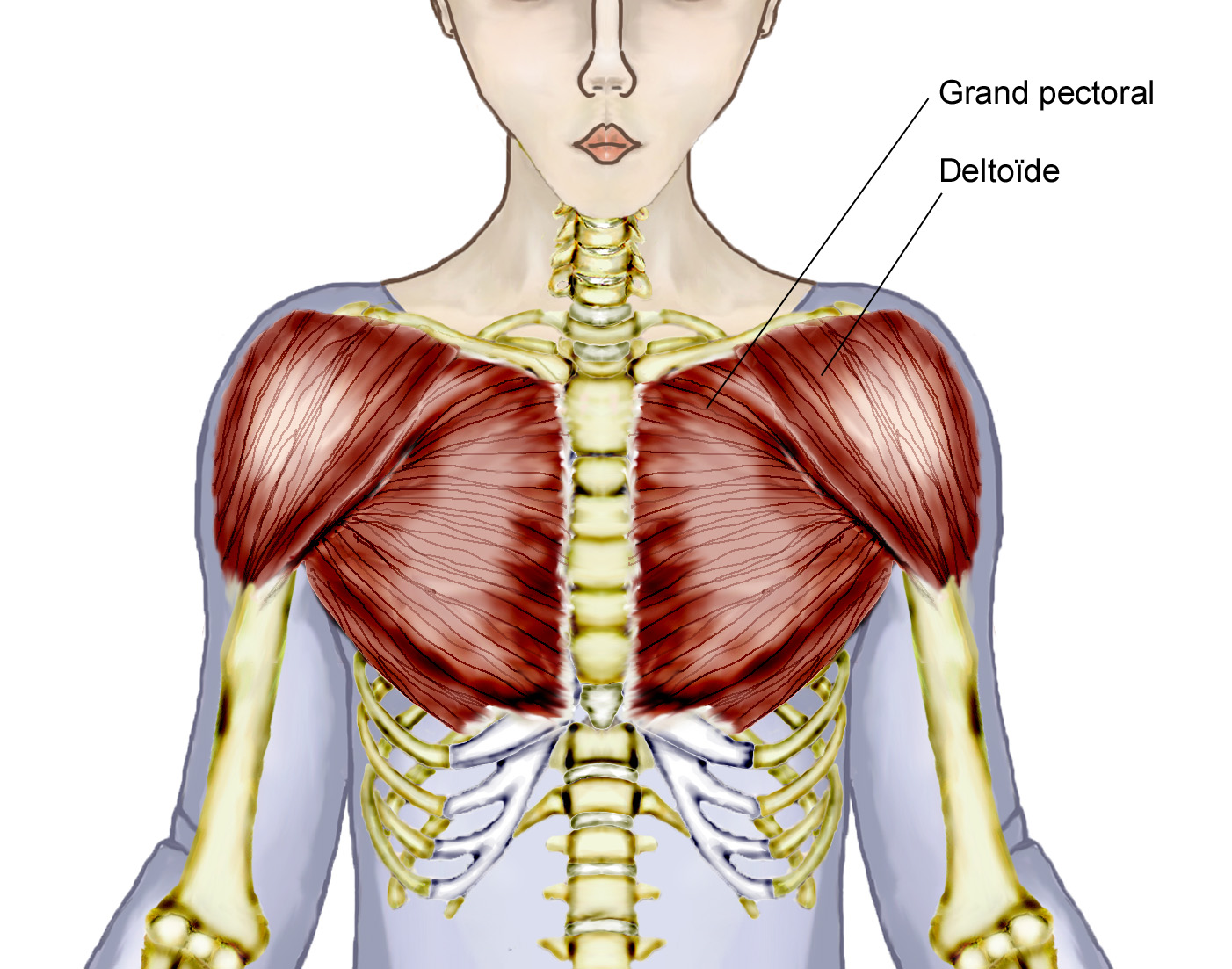
In this image, you will find part of the pectoral muscles mainly used in it. Start studying chest muscles anatomy. Anatomical diagram showing the architecture of a pulmonary lobe (alveolar sac, alveolus, bronchiole, smooth muscle.) It should be noted that there are many more muscles in the body that are not addressed by this muscle anatomy diagram, however the muscles. Human anatomy and physiology diagrams:
Overview Of Chest Muscles from www.modernheal.com In this post, you will learn the chest muscles anatomy which is easy since there are not so many muscles. Human anatomy diagram shoulder anatomy shoulder muscles shoulder muscles and chest. Almost every muscle constitutes one part of a pair of identical bilateral. Muscles, connected to bones or internal organs and blood vessels, are in charge for movement. All about the chest muscles. Attached to the bones of the skeletal system are about 700 named muscles that make up roughly half of a person's body weight. Meet your pectoralis major and pectoralis minor. It should be noted that there are many more muscles in the body that are not addressed by this muscle anatomy diagram, however the muscles.
Related posts of chest muscles diagram.
The primary function is certainly to provide support to the skeletal system and to facilitate its movements. The muscles of the intermediate muscle layer of the back are positioned beneath the trapezius and the latissimus dorsi. Their main function is contractibility. The drawings here present idealized versions of male and female torsos. Learn about each muscle, their locations & functional anatomy. Related posts of chest muscles diagram. Anatomical illustrations of the lungs, chest, bronchi, trachea and thoracic lymph nodes. Name and locate major muscles of the human body on a torso or diagram. The pectoralis minor muscle (not shown in the diagram) is located underneath the pectoralis major muscle, attaching to the coracoid. The gastrocnemius and soleus muscles taper and merge at the base of the calf muscle. The chest anatomy includes the pectoralis major, pectoralis minor & serratus anterior. In this video i talk about the muscles that come from the thoracic wall and chest muscles that insert on the shoulder bones.✅. Tough connective tissue at the bottom of the calf muscle merges with the achilles tendon.
Anatomical illustrations of the lungs, chest, bronchi, trachea and thoracic lymph nodes chest muscles diagram. To further your learning on the anconeus and arm arm anatomy in general check out the following article.


0 Komentar